Protect your central vision with expert care for age-related macular degeneration. Eye Surgeons of Indiana offers advanced treatments and personalized guidance to help preserve sight and improve quality of life.


What Is Age-Related Macular Degeneration?
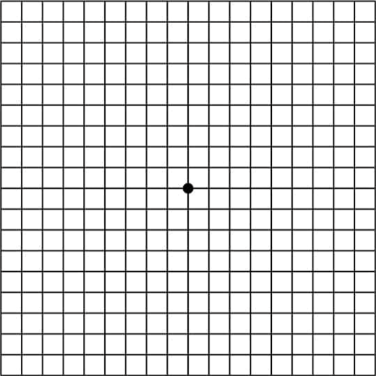
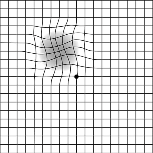
AMD is a progressive eye condition that affects the macula, the small central portion of the retina responsible for sharp, detailed vision. Damage to the macula can make it difficult to read, recognize faces, drive, or see fine detail. There are two main types of AMD.